 CHICAGO, Nov. 29 2009 The Royal Philips Electronics announced its response to an industry need by showing its portfolio of products designed to manage dose radiation at this year’s 95th annual meeting of the Radiological Society of North America (RSNA) in Chicago. New this year from Philips are several advancements and upgrades focused on redefining imaging, managing dose radiation and streamlining workflow to expand upon clinical capabilities. Philips will also showcase the company’s financing solutions for customers exploring radiology investments.
Advancements in medical imaging are continuously opening new doors in early diagnosis and disease management. Despite the progress of this technology, both the radiology and patient communities have concerns regarding the potential risks of radiation dose. As healthcare guidelines continue to shift and more intense focus is directed to dose radiation risk and reduction, it is increasingly important radiologists have access to imaging systems that may limit exposure while upholding image quality and ease of use.
“Hospitals are under intense pressure to deliver high-quality care while lowering costs and exceeding standards for patient safety,” said Gene Saragnese, CEO, Imaging Systems, Philips Healthcare. “Philips is dedicated to supporting our customers with systems that drive value and technologies that help enable a reduction of radiation dose to patients and hospital staff, while maintaining exceptional image quality to aid in the diagnosis and treatment of patients.”
New solutions to redefine imaging and manage dose for patients and clinicians, alike
iDose for Philips CT
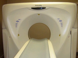
 Philips is unveiling a new X-ray dose reduction technique, iDose. The newest addition to Philips’ DoseWise suite of radiation management tools, iDose focuses on lowering X-ray dose while maintaining high image quality and fast reconstruction times. The iDose reconstruction technique, RapidView IR console, enables 20 times faster reconstruction than current hardware and lowers X-ray dose up to 80 percent while maintaining diagnostic image quality. As an ongoing commitment to streamlining workflow for radiologists, iDose was designed to provide the same look and feel of the original, higher-dose radiation images, so there’s no additional training needed. iDose is available for new and installed base Brilliance iCT and 64-channel scanners with expected delivery in the second half of 2010.
DoseAware
For interventional radiologists, Philips will introduce DoseAware, the latest addition to Philips’ DoseWise portfolio of solutions to help track X-ray exposure of patients and staff. DoseAware delivers real-time visualization, display and tracking of clinical staff X-ray dose to help healthcare professionals performing potentially lengthy interventional procedures manage their radiation exposure on the spot.
EasyDiagnost Eleva
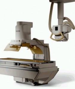
 A single fluoroscopy room is not cost-effective in today’s environment with a shift of exams to other modalities. The combination of Philips’ high-end digital vertical stand (or digital in-table detector) and the EasyDiagnost Eleva with ceiling suspension manages both high-quality digital radiography as well as fluoroscopy (DRF) applications, enabling users to perform gastro-intestinal work, as well as chest and skeletal exams, in just one room.
In addition, the DRF design consists of various single features to help enable lower X-ray doses to be delivered, including pediatric fluoroscopy settings, Grid Controlled Fluoroscopy and dose measurement. This is especially relevant in the pediatric setting.
The Eleva is easy to operate, fast and offers the ability to customize protocols and techniques for individual physician preferences. Based on open standards, the EasyDiagnost Eleva can seamlessly integrate into any hospital network.
GEMINI TF PET/CT
Showcasing Philips’ newest innovation in oncology care, the Philips GEMINI TF PET/CT streamlines the processes involved in staging, planning, therapy and follow-up in oncology care. With Generation 3 Time-of-Flight technology, the GEMINI TF system is the company’s fastest ever PET scanner, capturing data in less than 500 picoseconds, without compromising image quality or attention to dose radiation efficiency.
Philips GEMINI TF is seeing increased traction for oncology diagnosis and follow-up. The U.S. Centers for Medicare and Medicaid Services (CMS) final decision expands coverage for Medicare patients for 11 cancer types and adds new coverage for initial treatment in seven other cancer indications. FDG-PET exams, such as those performed on the GEMINI TF, can be performed as part of the initial treatment strategy and to further guide subsequent treatment strategies. Philips GEMINI TF family will now play an even larger role in women’s care due to the recent CMS decision to expand Medicare coverage for the reimbursement of exams for the initial staging of patients with biopsy-proven cervical cancer.
BrightView XCT SPECT/CT
Philips is showcasing new clinical images from the BrightView XCT. BrightView XCT using Astonish image reconstruction technology provides 50 percent better spatial resolution in cardiac SPECT studies compared to SPECT studies using filtered back projection. BrightView XCT helps enable low patient X-ray dose levels, high-resolution localization and high-quality attenuation correction with the potential for fewer artifacts and shorter exam times.
Additionally, Philips will highlight new clinical evidence that Philips’ Astonish algorithms for nuclear cardiology help clinicians improve their diagnostic accuracy and increase their diagnostic confidence.
Allura Xper FD20 Release 7
For new and existing customers, the new release of the Allura Xper FD20 combines a full range of advanced interventional tools and seamless multi-modality cath lab integration. It also offers a balance between superb image quality and low X-ray dose during lengthy procedures. The system is upgradable, offering access to the latest clinical and service innovations as part of the lifetime Philips commitment.
Following the upgrade, the Allura Xper system will have advanced tools to support image-guided procedures. The MR/CT Roadmap feature allows clinicians to synchronize live fluoroscopy with previously Acquired MRA/CTA datasets to help reduce contrast media use and X-ray dose compared to standard 2D or 3D navigation.
